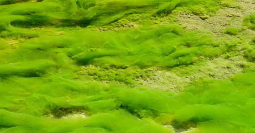
Protothecosis is a rare infection due to an achlorophyllous unicellular alga, Prototheca, which is ubiquitous in nature. It causes diseases in both animals and humans. It may colonize human skin, the respiratory tract, and the gastrointestinal tract. Cases of human protothecosis have been reported worldwide. Between 1964, when the first case report was published, and November 2012, there have been slightly more than 160 cases of human protothecosis reported in the literature. Among Prototheca species, Prototheca wickerhamii is the most common cause of human infections, followed by Prototheca zopfii.
The 3 most recognized forms of human protothecal infections are cutaneous (72%), olecranon bursitis (18%), and disseminated systemic infections (10%). Localized infections (cutaneous and olecranon bursitis) occur more frequently in immunocompetent individuals, whereas disseminated infection tends to be seen in immunocompromised patients, in whom disseminated systemic infection can be life-threatening.
Cutaneous infections are frequently preceded by minor trauma or surgical wounds exposed to soil or water but may occur without clear prior compromise of skin / mucosa. The typical cutaneous manifestation is a painless, slowly progressive, well-circumscribed plaque or papulonodular lesion that may become eczematoid or ulcerated. A variety of lesion types have been reported, however, including erythematous plaques, pustules, papules, vesicles, ulcers, synovitis, and chronic draining wounds.
Protothecal olecranon bursitis, due to inoculation after trauma or wound contamination, presents as tenderness, erythema, and serosanguineous bursal discharge several weeks after inoculation / infection.
Immune deficiency is the major risk factor for severe systemic disease involving skin and subcutaneous tissue, blood, meninges, heart valves, lymph nodes, lung, intestines, gall bladder, liver, spleen, and peritoneum. In immunocompromised hosts (hematological diseases, solid organ transplantation, cancers, chronic steroid use, diabetes mellitus, autoimmune diseases, HIV/AIDS), localized cutaneous infections may disseminate and progress to systemic algaemia. At the time of this writing, 19 reported cases of proven systemic Prototheca algaemia have been published in the English language literature. Significant cholestatic jaundice from granulomatous hepatitis in patients with disseminated deep visceral protothecal algaemia has been reported.
In patients with Prototheca algaemia, the source of infection is only identified in 50% of patients, of which central venous catheters and cutaneous lesions are the most common nidi of algaemia. In central venous catheter-associated Prototheca algaemia, concomitant polymicrobial bacteremia (most commonly Enterococcus faecalis, vancomycin-resistant E. faecium [VRE], Escherichia coli, and Candida glabrata) has been observed.
